Understanding Mammograms
According to experts, a mammogram is the best screening tool for detecting breast cancer at its earliest, most treatable stages 1. Using special x-ray imaging, a mammogram can reveal both harmless and cancerous growths even when they are too small to be felt through a breast self-exam or clinical breast exam.

What to Expect of a Mammogram
A mammogram can be performed in a variety of settings, including hospital clinics, mobile vans or your doctor’s office. It involves standing or sitting in front of a specialized x-ray machine. A technologist will position your breast on a platform. A clear plastic plate will press the breast flat, enabling the maximum amount of breast tissue to be examined with the least radiation possible.
The compression of the plates may cause some minor discomfort, but no significant pain. If you feel pain, you should inform the technologist immediately. If you are menstruating, you may want to schedule your mammogram appointment a week after your period ends when your breasts are not swollen or tender. And if you have breast implants, you should tell your technologist so extra care can be taken to prevent rupturing the implant.
During the mammogram procedure, you will need to remove your clothes from the waist up. For this reason, you may want to wear loose clothing and a shirt that you can take off easily. You will also want to refrain from wearing any deodorant, lotions or powders. These substances can alter the x-ray quality.
Many mammogram facilities now use skin markers to help identify areas of concern or normal breast variations such as surgical scars and raised moles. These adhesive markers are placed on the breast skin and can be seen on your mammogram images. This alerts the radiologist and enables a fast accurate reading of the mammogram film.
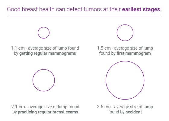
Mammogram Results
After your mammogram is completed, a radiologist will examine the films and determine if there are breast abnormalities that need further exploration. If your mammogram results are not back within 10 days, contact your doctor or the mammogram facility. Don’t assume that the results are normal just because you haven’t received them. If the mammogram shows a breast abnormality, talk with your doctor about the appropriate next steps.
There are two basic types of mammograms, screening and diagnostic. Participating in screening mammograms can detect tumors at their earliest stages.
https://www.fda.gov/consumers/consumer-updates/mammography-what-you-need-know
Screening Mammograms
This type of mammogram is used for women who are asymptomatic, meaning they have no complaints or symptoms of breast cancer. Its purpose is to identify breast abnormalities before they can be detected through breast self-exams or clinical breast exams. Screening mammograms generally involve taking x-rays from two views of each breast and can be completed in less than 15 minutes. According to the FDA, screening mammograms can detect 85% to 90% of all breast cancers.
Diagnostic Mammograms
A diagnostic mammogram is administered to women who are experiencing symptoms of breast cancer or have detected a breast abnormality. It involves a more thorough examination and is used to determine the exact size and location of the breast abnormality. Diagnostic mammograms generally take several x-ray views of the breast and require approximately 30 minutes to complete. Women having follow up imaging or a personal history of breast cancer will usually require a diagnostic mammogram.
3D mammography or Digital breast tomosynthesis (DBT)
During a 3D mammogram, the breast is positioned and compressed the same way as a 2D mammogram, but the X-ray tube moves in an arc over the breast. This captures multiple images and allows for these projections to be “synthesized” by a computer. A radiologist can then view the breast in small 1 millimeter slices with greater detail because this reduces the overlap of tissue you see on a 2D mammogram. Compared to standard 2D mammography, 3D mammography has been found to increase the number of cancers detected and to decrease the number of false-positive examinations.
Check with your insurance provider to see if 3D mammography is covered.